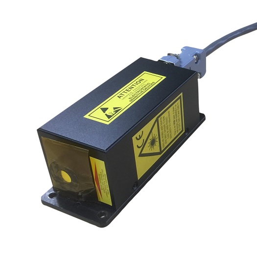
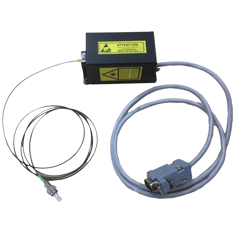
Pavilion Integration WhisperIT® laser modules and Lapis® multi-laser engines with customized collimation beam shaping are being used to reduce phototoxicity, and improve imaging speeds 100-1000x point scanning methods.

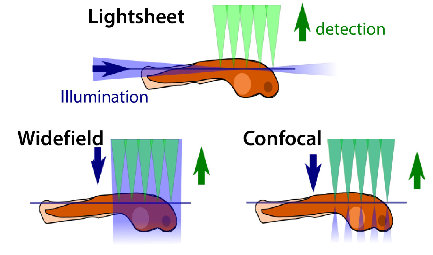
Light sheet microscopy distinguishes itself from more basic light microscopy techniques by only exciting fluorophores within its focal plane, greatly reducing potential phototoxicity levels and allowing for high spatial and temporal resolution.
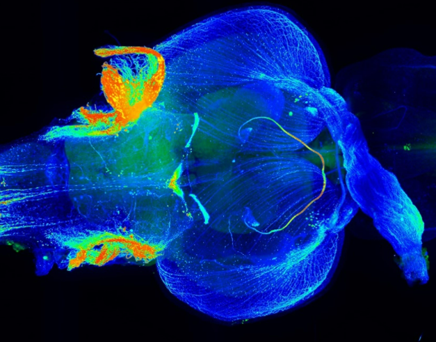
Light-sheet fluorescence microscopy (LSFM) uses a thin sheet of light to excite only fluorophores within the focal volume. Light sheet microscopes (LSMs) have a true optical sectioning capability and, hence, provide axial resolution, restrict photobleaching and phototoxicity to a fraction of the sample and use cameras to record tens to thousands of images per second. LSMs are used for in-depth analyses of large, optically cleared samples and long-term three-dimensional (3D) observations of live biological specimens at high spatio-temporal resolution.

Because light sheet fluorescence microscopy scans samples by using a plane of light instead of a point (as in confocal microscopy), it can acquire images at speeds 100 to 1,000 times faster than those offered by point-scanning methods. Users benefit from custom collimation beam shaping to ensure illumination uniformity.


